Upper Leg Tendon Anatomy - The Successful Treatment of Achilles Tendon Issues in Dogs ... : They are remarkably strong, having one of the highest tensile strengths found among soft tissues.
Upper Leg Tendon Anatomy - The Successful Treatment of Achilles Tendon Issues in Dogs ... : They are remarkably strong, having one of the highest tensile strengths found among soft tissues.. Collectively, they act to dorsiflex and invert the foot at the ankle joint. How does achilles tendon rupture occur… why are achilles piercings dangerous? This article will discuss the anatomy and function of the achilles tendon. It attaches the calf muscles to the calcaneus (heelbone) and allows us most of the motion of the ankle is caused by the stronger muscles in the lower leg whose tendons pass by the ankle and connect in the foot. The pads of the machine are situated at the achilles tendon.
It is formed when the soleus muscle tendon joins with the gastrocnemius tendon. Leg anatomy muscles and tendons how to fix achilles. Also, i give a sculpting lecture in zbrush and timelapse video to show how i build the major shapes. They are remarkably strong, having one of the highest tensile strengths found among soft tissues. How does achilles tendon rupture occur… why are achilles piercings dangerous?
Appendicular Muscles of the Pelvic Girdle and Lower Limbs ... from pressbooks-dev.oer.hawaii.edu This mri wrist coronal cross sectional anatomy tool is absolutely free to use. 1280 x 1520 jpeg 166 кб. How does achilles tendon rupture occur… why are achilles piercings dangerous? Collectively, they act to dorsiflex and invert the foot at the ankle joint. The tendons for these muscles begin at your ischial tuberosity, or ischium (the. Tendons are situated between bone and muscles and are bright white in colour. This may result in tendon subluxation; An anatomical and biomechanical study.
What are the functions of patella.
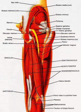
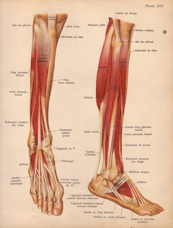
The calcaneal tendon, also known as the tendon of achilles, is a posterior leg tendon — a fibrous connective tissue that joins muscles in the back of the leg. Tendons are fibrous cords attached to muscles and bone. Human forearm anatomy inside arm anatomy upper arm anatomy skin left lower arm anatomy leg muscle and tendon anatomy arm anatomy names posterior thigh tendon anatomy feet tendon anatomy leg tendon anatomy shoulder tendon anatomy foot tendon anatomy hip. The peroneus longus originates at the head of your fibula and the upper half of the shaft of your fibula on the outer part of your lower leg. Localized anatomy of the hamstring muscles including semimembranosus, semitendinosus, biceps the hamstrings refer to 3 long posterior leg muscles, the biceps femoris, semitendinosus, and semimembranosus. Common tendon of superficial posterior leg muscles; Originates from the lateral condyle of the tibia and the medial surface of the fibula. The lower leg is comprised of two bones, the tibia and the smaller fibula. ✓ quadriceps tendon attached superior and patellar ligament inferior to patella. Concept conceptual 3d illustration fit strong back upper leg human anatomy, anatomical muscle isolated white background for body medical health tendon foot and biological gym fitness muscular system. Lie prone on a hamstring curl machine. Tendon, tissue that attaches a muscle to other body parts, usually bones. Hands are outstretched, holding onto the handles of the bench.
There is no real division between the core and the upper leg; Also, i give a sculpting lecture in zbrush and timelapse video to show how i build the major shapes. Related posts of muscle anatomy upper leg. What are the functions of patella. Leg anatomy muscles and tendons how to fix achilles.
One More Plate: How the Hamstrings Work from 3.bp.blogspot.com The lower leg is comprised of two bones, the tibia and the smaller fibula. The human leg, in the general word sense, is the entire lower limb of the human body, including the foot, thigh and even the hip or gluteal region. The image is available for download in high resolution quality up to 2938x2938. It is formed when the soleus muscle tendon joins with the gastrocnemius tendon. Common tendon of superficial posterior leg muscles; Plantar flexion of the foot, ankle joint stabilizer. Leg anatomy muscles and tendons how to fix achilles. Lie prone on a hamstring curl machine.
Thompson's test, achilles tendon rupture.
The tendons that control movement in your hands, wrists and fingers run through your forearm. Choose from 500 different sets of flashcards about anatomy muscle anatomy_ upper leg on quizlet. Originates from the lateral condyle of the tibia and the medial surface of the fibula. Tendons are fibrous cords attached to muscles and bone. .16 penile numbness and perineum tenderness.18 any suggested exercises or stretches?.22 leg musculature 209 elbow tendonitis and saddle sores. What are the functions of patella. 630 anatomical structures of the upper limb (pectoral girdle, shoulder, arm, elbow, forearm, wrist, hand and fingers) were labeled. This mri wrist coronal cross sectional anatomy tool is absolutely free to use. Thompson's test, achilles tendon rupture. Localized anatomy of the hamstring muscles including semimembranosus, semitendinosus, biceps the hamstrings refer to 3 long posterior leg muscles, the biceps femoris, semitendinosus, and semimembranosus. Lie prone on a hamstring curl machine. ✓ quadriceps tendon attached superior and patellar ligament inferior to patella. The human leg, in the general word sense, is the entire lower limb of the human body, including the foot, thigh and even the hip or gluteal region.
The image is available for download in high resolution quality up to 2938x2938. Use the mouse scroll wheel to move the images up and down alternatively use the tiny arrows (>>) on both side of the image to move the images. This mri wrist coronal cross sectional anatomy tool is absolutely free to use. Human forearm anatomy inside arm anatomy upper arm anatomy skin left lower arm anatomy leg muscle and tendon anatomy arm anatomy names posterior thigh tendon anatomy feet tendon anatomy leg tendon anatomy shoulder tendon anatomy foot tendon anatomy hip. The calcaneal tendon, also known as the tendon of achilles, is a posterior leg tendon — a fibrous connective tissue that joins muscles in the back of the leg.
Items similar to 1905 leg muscles, tendons & ligaments ... from i.etsystatic.com Muscle/tendon inflammation and pain along anterio… Leg muscle anatomy chart | amulette. Hands are outstretched, holding onto the handles of the bench. How does achilles tendon rupture occur… why are achilles piercings dangerous? Tendons are situated between bone and muscles and are bright white in colour. The pads of the machine are situated at the achilles tendon. Collectively, they act to dorsiflex and invert the foot at the ankle joint. Horse leg basic anatomy tendons подробнее.
Originates from the lateral condyle of the tibia and the medial surface of the fibula.
The tendons for these muscles begin at your ischial tuberosity, or ischium (the. Hands are outstretched, holding onto the handles of the bench. It is formed when the soleus muscle tendon joins with the gastrocnemius tendon. Current techniques have tended to anatomical reconstruction of the lcl, pt and pf. This may result in tendon subluxation; Collectively, they act to dorsiflex and invert the foot at the ankle joint. Related posts of muscle anatomy upper leg. There are four muscles in the anterior compartment of the leg. The lower leg is comprised of two bones, the tibia and the smaller fibula. This mri wrist coronal cross sectional anatomy tool is absolutely free to use. Tendons are fibrous cords attached to muscles and bone. An anatomical and biomechanical study. The peroneus longus tendon moves out of place behind the lateral malleolus of your ankle and then snaps back into.
Share this post
0 Response to "Upper Leg Tendon Anatomy - The Successful Treatment of Achilles Tendon Issues in Dogs ... : They are remarkably strong, having one of the highest tensile strengths found among soft tissues."


0 Response to "Upper Leg Tendon Anatomy - The Successful Treatment of Achilles Tendon Issues in Dogs ... : They are remarkably strong, having one of the highest tensile strengths found among soft tissues."
Đăng nhận xét